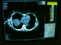
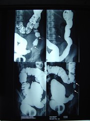
CT Guided percutaneous needle biopsy of pulmonary masses

CT

Percutaneous apiration of a pulmonary tumor can be performed through the chest wall, using CT scanning to guide the placement of the needle within the lesion. The yield froma a percutaneous approach is generally higer than the yield from fiberoptic bronchoscopy. Overall, the diagnostic yield from percutaneous needle aspiration has been reported to range from 43 to 97 percent

Gerardo Morales, M.D.

technorati tags: , , , , , ,

A rare thoracic tumor

Useful links:

50 y/o male with a history of one year with respiratory symptoms, he was diagnosed treated at Guatemala where he lives. Now he comes to Mexico City seeking for a second opinion and other options of therapy. At the physical examination he is well, has diminished breath sounds and just sinus tachycardia.

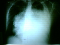
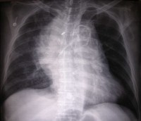
Here is his chest x ray:
Chest x-ray

What’s the diagnosis?

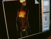
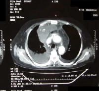
A PET-CT was done:
PET-CT

What’s your diagnosis?

Thymic Carcinoma

Thymic carcinoma displays distinct morphology and biology, different in many ways from thymoma. It is composed of highly atypical cells with cytoarchitectural features of carcinoma similar to those seen in other organs. Although many lymphocytes can be seen in its stroma, they are of B cell type and mature T cell type; thymic carcinoma lacks the immature T cell lymphocytes of thymoma.
Thymic carcinoma is classically not associated with paraneoplastic syndromes such as myasthenia gravis. However, one study reported 22 cases of primary thymic epithelial neoplasms which showed combined features of thymoma and thymic carcinoma; the carcinoma and thymoma were either found synchronously, or carcinoma developed with a preexisting thymoma after 10 to 14 years.
Incidence Primary thymic carcinoma is a rare neoplasm. At the National Cancer Center of Tokyo, 17 cases were resected over a 29 year period, during which time 79 thymomas and 2500 lung cancers were treated.
Clinical and radiographic manifestations Primary thymic carcinoma is a tumor of adulthood (mean age of 46 to 54 years). Most patients complain of chest symptoms, including pain, cough, superior vena cava syndrome, or general symptoms such as fatigue, weight loss, anorexia; fewer than 10% of patients are asymptomatic. Chest radiographs and computed tomography show an anterior mediastinal mass infiltrating along the pleura or mediastinum, with frequent necrosis or rare calcifications.

technorati tags: , , , , , , , , , , , , , ,

Another Tension-type Pneumothorax

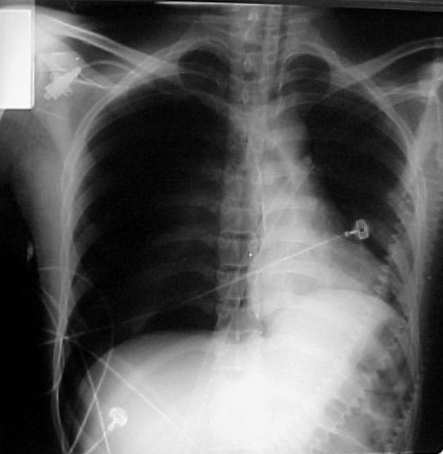
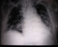
Here is another chest x-ray that shouldn’t have been taken:

Tension-type Pneumothorax

As reference, you can trackback this earlier post.

Regards,

Jon Mikel Iñarritu, M.D.

technorati tags: , , , , , , , , , ,

Open Fractures Classification and its clinical manifestations

Open fractures are surgical emergencies because of its complications (soft tissue damage, infection, hemorrhage).

We at emergency department need a classification system that it is intented to determine the degree of that damage.

We have to make a rapid (also a good) interrogation of lession mechanisms (what, when,  where, who, associated lessions). REMEMBER: If a low-grade open fracture has 6hrs from the begining, it’s considered infected.

On 1984, Gustilo and Anderson made a classification system, wich is the following:

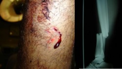
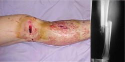
Type I open fracture. Wound less than 1 cm, without contamination and minimal injury of soft tissue.
Type I open fracture - 1
Type I open fracture - 2


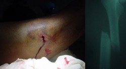
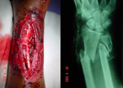
Type II open fracture. Wound between 1 and 10 cm, mild contamination, extensive soft tissue damage and minimal to moderate crushing component.
Type II open fracture - 1
Type II open fracture - 2

Type III-A open fracture. Wound larger than 10 cm, severe contamination and severe crushing component.

Type IIIA open fracture

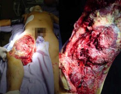
Type III-B open fracture. Wound larger than 10 cm, severe contamination and severe loss of tissues.
Type IIIB open fracture - 1
Type IIIB open fracture - 2

Type III-C open fracture. Wound larger than 10 cm, severe contamination and neurovascular injury.
Type IIIC open fracture - 1
Type IIIC open fracture - 2

REFERENCES:
Gustilo, R.; Mendoza, R.; Williams, D. Problemsin the management of type III (severe) open fractures: A new classification of typeIII open fractures. J Trauma 24:742, 1984.

Regards,
Jon Mikel Iñarritu, M.D.

technorati tags: , , , , , , , , , , , , ,

Cardiac Tamponade

Physical findings & Clinical Presentation:

• Severeconstant pleuritic pain that localizes over the anterior chest and may radiateto arms and back; it is differentiated from myocardial ischemia, becausethe pain intensifies with inspiration and is relieved by sittingup and leaning forward.
• Pericardial friction rub is best heard with patientupright and leaningforward and by pressing the stethoscope firmly against the chest; itconsists of three short, scratchy sounds:
• Signs and symptoms:

  1. Dyspnea
  2. Orthopnea
  3. Interscapular pain
  4. Tachycardia
  5. Refractary hypotension
  6. Distended neck veins
  7. Paradoxicalpulse
  8. Chest x-ray: Cardiomegaly, clear lungs (may be normal)
  9. ECG: Low amplitude of QRS, electrical alternans

Treatment:
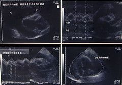
Immediatepericardiocentesispreferably by needle paracentesis with the use of echocardiography, fluoroscopy,orCT Aspirated fluidshould be sent for analysis (protein, LDH, cytology, CBC, Gram stain, AFBstain) and cultures for AFB, fungi, and bacterial C&S.

•

tamponade

Cardiac Tamponade

tamponade

Cardiac Tamponade

Regards,
Jon Mikel Iñarritu, M.D.

technorati tags: , , , , , , , , , ,

Traumatic mediastinal hematoma

A 64 years old politraumatized male, arrive to ER department directly to trauma room. He was on shock.

  • Refractary hypotension
  • Tachycardia and tachypnea
He had no signs of tamponade.
Here we have the images (best viewed in large size):

Initial chest x-rayCT scan

Post-surgery image (best viewed in large size):

Post-op chest x-ray

The final diagnosis was Traumatic Mediastinal Hematoma.
You can notice esophagus displacement and complications of filling cardiac cavities.
Regards,
Jon Mikel Iñarritu, M.D.

technorati tags: CXR, mediastinal hematoma, CT scan, Clinical cases, medicine, mediastinum, unbounded medicine, chest x-ray, x-ray, xray

An extremely rare case

22 year old female with painless slowly progressive, ophtalmoplegia with diplopia and ataxia.

On physical examination we found this:

  • Short stature.
  • Arrhythmic heart sounds.
  • Bilateral ptosis.
  • Ophtalmoplegia.

Ptosis & ophtalmoplegia

Bilateral fundoscopy examination:

Eye Another Eye

A characteristic bilateral pigmentary retinosis.

An EKG was performed:

1st degree AV block
A first degree AV block was found on this patient.

Then we ordered a lumbar punction to chemical and cytologic analysis.

The cerebrospinal fluid was cloudy with no other anormalities except elevated proteins (>100mg/dL).

With this data now we can think about a chronic progressive external ophtalmoplegia.

Differential diagnosis should include:

  1. Isolated CPEO
  2. Kearns-Sayre syndrome
  3. Oculopharyngeal muscular dystrophy
  4. Myotubular myopathy
  5. Myotonic dystrophy
  6. Oculopharyngeal muscular dystrophy

The next step in the diagnosis of this case is to perform an electromyography (myophatyc pattern), electrorretinography (slow transmission) and a muscle biopsy.

The diagnosis is:

Kearns-Sayre syndrome wich is a mitochondrial cytopathy that is characterized by CPEO, retinal pigmentary changes, and heart block. Patients are typically normal at birth. Progressive ophthalmoplegia usually develops between 5 and 20 years of age, although it may occur earlier. Most cases are sporadic.

The ocular findings include bilateral and symmetric involvement of the horizontal and vertical muscles, bilateral ptosis, and normal pupils. An atypical pigmentary retinopathy (“salt and pepper”) may be present; some patients have a corneal opacity. The ophthalmoplegia progresses slowly over many years and is often asymptomatic because it is insidious and bilaterally symmetric. As the extraocular myopathy progresses, generalized muscle weakness and other systemic manifestations may occur.

Nonocular manifestations include:

  • Cardiac — Heart block, sometimes even sudden death (may require monitoring with sequential electrocardiograms or treatment with a pacemaker) [2,3]
  • Neurologic — Deafness and vestibular dysfunction, cerebellar ataxia, corticospinal dysfunction, electroencephalogram abnormalities, elevated cerebrospinal fluid protein (>100 mg/dL), or widespread muscular dystrophy
  • Endocrine and metabolic — Short stature.

Images and clinical case information provided by: Dr. Arturo Ramírez M. from Mexico City.

Thank you for your support, Arturo.

Regards,

Jon Mikel Iñarritu, M.D.

technorati tags: , , , , , , , , ,

RA & Pulmonary disease

A 76 years old female comes to the emergency department because shortness of breath. She has been postrated since 2 months ago, when she wasn’t able to move her legs anymore. Shortness of breath since 1 week ago (NYHA IV). She states that she has “rheumas”. She gives no other information. She has been taking methotrexate since 4 years ago, in interval periods of time, she doesn’t know which doses. On physical examination you note a postrated elder woman with severe physical deformities. You also found this:

general appearence deformity

clubbing swan neck

cxr lat cxr

This is an extremely advanced case of rheumatoid arthritis. But, what associations with pulmonary disease does RA have? What kind(s) of pulmonary disease(s) does this patient have (or could have)?