Do not explode a firecracker in your hand never (update)

BEWARE! Gory Images

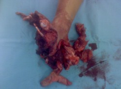
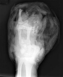
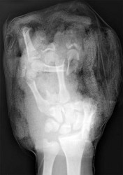
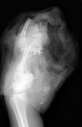
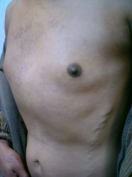
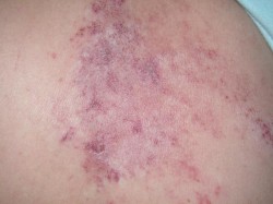
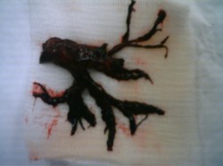
Another case of firecracker associated injuries here.

firecracker1

firecracker2

For those who don’t believe in that this kind of accidents really happen, this is a story who GruntDoc wrote, with the same outcome.

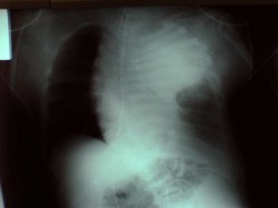
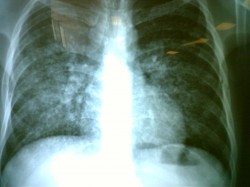
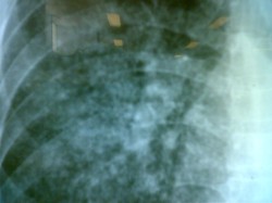
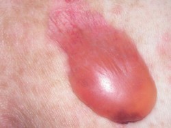
The pictures are self explaining.

GruntDoc says: “The devastation on this X-ray is nothing compared to the actual flesh-and-blood injury. A life changed in a second.”

Traumatic thoracic aortic rupture

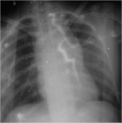
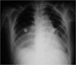
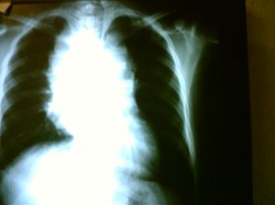
A previously healthy 17 yo woman came in to the ER because she has suffer an automovilistic accident. On the initial evaluation she was with pulse ‘parvus et tardus’ with altered mental status. A chest x-ray was ordered and found an aortic rupture.
Initial Chest X-Ray

Because the bleeding was contained and she has just a little hemodynamic instability, the choice was endovascular treatment of traumatic thoracic aortic rupture.
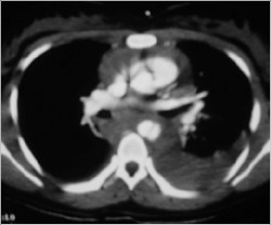
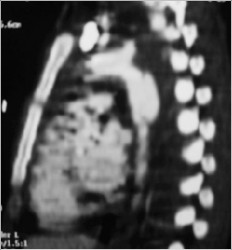
A CT Scan was ordered preoperatory:

CT Scan - Aortic Rupture

CT Scan - Aortic rupture

Angio TC

Endovascular Procedure

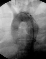
Flouroscopy - Stent

After Procedure

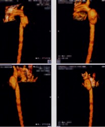
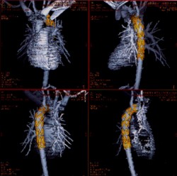
Reconstruction

Postoperatory Chest X-ray

The outcome was excellent and the patient is in good shape rightnow with a normal lifestyle.

Regards,

Jon Mikel Iñarritu, M.D.

Insulinoma & Whipple triad – Surgery (update)

Insulinoma

Insulinoma is the most common functioning tumor of the pancreas, and affected patients present a tableau of symptoms referable to hypoglycemia (symptoms of catecholamine release), mental confusion and obtundation. Many patients have symptoms for years. Some have been greatly troubled by emotional instability and fits of rage, often followed by somnolence.

Incidence: This tumors are so rare. Probably the best series of insulinomas is from the Olmsted County (Mayo Clinic). This study (cohort) was seen over a sixth decade period (’27 to ’86). The incidence there was 0.4 per 100,000 person-years (or four cases per million per year).

The diagnostic hallmark of the syndrome is the so-called Whipple triad, wich consists in symptoms of hypoglycemia (catecholamine release), low blood glucose level (40 to 50 mg/dL), and relief of symptoms after intravenous administration of glucose. The triad is not entirely diagnostic, because it may be emulated by factitious administration of hypoglycemic agents, by rare soft tissue tumors, or occasionally by reactive hypoglycemia. The clinical syndrome of hyperinsulinism may follow one of two patterns or sometimes a combination of both.

The symptom complex may be due to autonomic nervous overactivity, expressed by fatigue, weakness, fearfulness, hunger, tremor, sweating, and tachycardia, or alternatively, a central nervous system disturbance with apathy (or irritability or anxiety), confusion, excitement, loss of orientation, blurring of vision, delirium, stupor, coma, or convulsions.

Because cerebral tissue metabolize only glucose, prolonged profound hypoglycemia may cause permanent brain damage. Clinicians need to be alert to this when attempting to induce hypoglycemia by fasting.
Preoperative NPO orders must be accompanied by intravenous administration of glucose.

The diagnosis: Is established by demonstrating inappropriately high serum insulin concentrations during a spontaneous or induced episode of hypoglycemia. Then imaging techniques are used to localize the tumor. The available procedures include spiral CT, arteriography, ultrasonography (transabdominal and endoscopic), and 111-In-pentetreotide imaging. Transabdominal ultrasonography is our preferred initial test.

Insulinomas are small (usually smaller than 1.5 cm), usually single (only 10% are multiple and those are usually associated with MEN 1 syndrome), usually benign (only 5% to 10% are malignant), and usually hard to find. Success in localization often parallels the degree of invasiveness of the study.

Its treatment:
Treatment for insulinoma is surgical. At operation, the incision is dictated by operator preference, either a midline incision from the xiphoid to below the umbilicus or a bilateral subcostal incision. Exposure should be generous, and mechanical ring retractors are an asset. The entire abdomen should be explored, with particular attention being paid to possible liver metastases.

The head of the pancreas should be palpated carefully and examined anteriorly and posteriorly; the body and tail of the pancreas should be palpated, dividing any ligamentous attachments to the spleen, delivering the spleen into the wound, and rotating the tail anteriorly to allow palpation and visualization.

Anyone operating patients with pancreatic islet adenomas should be familiar with techniques for, and limitations of, intraoperative ultrasonography. The higher-resolution (7.5-to 10-MHz) transducers are used in the pancreas; because of its greater depth of penetration. Islet tumors are detected as sonolucent masses, usually of uniform consistency. Several reports attest to the high degree of accuracy of intraoperative ultrasound.

Review of current literature leads to the almost startling conclusion that nearly all syndromes of hyperinsulinism are due to insulinomas and that nearly all insulinomas can now be detected before or during surgery. Having said this, there appears to be scant justification for an empirical (“blind”) partial pancreatectomy except in patients with insulinoma plus MEN 1.

Most insulinomas are benign and can be enucleated. Nutrient vessels in the bed of the adenoma should be cauterized.

The 10% of patients with hyperinsulinism who have MEN 1 syndrome have multiple islet tumors, one of which is usually dominant and responsible for the excessive insulin output.
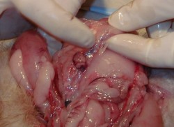
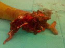
Insulinoma

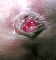
Thrombosed Hemorrhoid

Thrombosed Hemorrhoid

Thrombosed external hemorrhoids often occurs in grade III-IV internal hemorrhoids, and can cause excruciating pain, and patients will often present acutely. In such cases, surgical evacuation of the hemorrhoid can produce immediate relief.
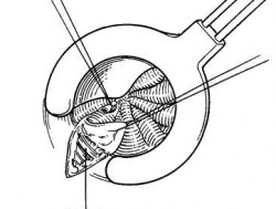
The treatment of choice is hemorrhoidectomy with partial lateral internal sphincterotomy, which consists in incised the mucosa on each side of the hemorrhoid and extended outward toward the anoderm. The anoderm and the hemorrhoidal mass are elevated off the transverse fibers of the internal sphincter muscle. Then the mucosa is approximated with continuous 3-0 absorbable sutures, leaving 2 to 3 mm of the anoderm left open for drainage.

hemorrhoidectomy

Regards,

Jon Mikel Iñarritu, M.D.

Link to: Hemorrhoidal Prolapse

Complications of subclavian venopuncture

This is a rare complication of subclavian venopuncture:

Inadvertent subclavian arterial puncture

Inadvertent subclavian arterial puncture

In inexperienced hands it could be a fatal procedure. So, in this case, it was too late when we try to stop that massive bleeding after a surgery interconsultation.

Regards,

Jon Mikel Iñarritu, M.D.

technorati tags: , , , , , ,

Pemphigus and bullous pemphigoid. 70 y/o woman with cutaneous lesions

This woman has this cutaneous lesions distributed all over the body. She has history of vitiligo.

Pemphigus and bullous pemphigoid.

These are autoimmune blistering diseases. They are caracterized by the production of antibodies directed against different epitopes, resulting in intraepidermal blistering in pemphigus and subepidermal blistering in pemphigoid. The term pemphigus comes from the greek, pemphix, that means bubble. It was used to describe most blistering diseases until 20th century.

Pemphigus
Pemphigus is a group of rare, chronic, autoimmune and potentially fatal diseases of mucous membranes and skin.Three types of pemphigus have been described:

Pemphigus vulgaris
Pemphigus foliaceus
Paraneoplastic pemphigus

Is caracterized by the production of autoantibodies, in pemphigus vulgaris IgG, against adhesion proteins. This alters the intercellular cement that holds epidermal cells together. Resulting in blister formation. Clinically is caracterized by flaccid bullae that tipically begin in the oropharynx and then may spread to involve the skin. With a predilection for the scalp, face, chest, axillae and groin.

Bullous Pemphigoid
Mostly afects persons 60 yeard of age and older. Pemphigoid refers to pemphigus-like blistering disease but is less aggressive than pemphigus vulgaris. It is also a chronic and recurrent disease of autoimmune origin. As in pemphigus vulgaris, the precise reason for the formation of autoantibodies is unkown. A small group of patient bullous pemphigoid is drug-induced. Penicillamine and furosemide are the most frequently implicated. A common presentation is a widespread blistering eruption in a middle age or eldery person who is taking multiple medication.

The case above was diagnosed as bullous pemphigoid.

Regards,

Gerardo Morales-Mora, M.D.