Cascade Stomach: An unsusual cause of abdominal pain

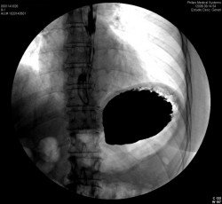
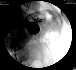
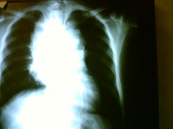
A woman came to the office because of sharp epigastric pain. On physical examination nothing was wrong except of epigastric pain. An endoscopy was performed and reported as normal. An Upper Gastrointestinal Tract Barium Examination was ordered and we found a cascade stomach, which is a rare finding. We exlude other causes of abdominal pain in the first place.

Here are the images.

Cascade Stomach 1

Cascade stomach 2

Cascade stomach 3

As you can see in this series, the fundus, still lies in its usual position relative to the structures of the left upper quadrant but the proximal portion of the body of the stomach is in an abnormally anterior and superior position. When this type of stomach is filled with barium (erect position), static roentgenograms may demonstrate a separate fluid level confined to the fundus. At fluonoscopy, barium first fills the dependent, posterior fundus to the highest level of the “ridge” and then spills or “cascades” into the body and antrum.

On the next day we have performed an esophageal manometry and the patients had lower esophageal incompetence, so we performed a laparoscopic Nissen fundoplication with gastropexia.

Now the patient is painless and in excellent condition.

Regards,

Jon Mikel Iñarritu, M.D.

Purpura fulminans as severe complication of meningococcal infection

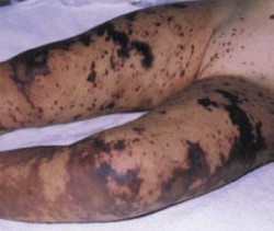
Purpura fulminans

It is a severe condition due to meningococcal sepsis, it occurs in 15 to 25% of those patients with meningococcemia.

The clinical picture is as follows: acute onset of cutaneous hemorrhage and necrosis secondary to vascular thrombosis and disseminated intravascular coagulation. Often there is pain followed by petechiae. Ecchymoses develop and evolve into painful indurated, well-demarcated purple papules with erythematous borders (as you can see in the image this lesions are coalescent). Then this lesions progress to necrosis with formation of bullae and vesicles. Gangrenous necrosis can follow with extension into the subcutaneous tissue and occasionally involves muscle and bone.

purpura fulminans

To prevent this complication of meningococcemia you have to be prompt and agressive with IV antibiotics and support of vascular perfusion. The use of Xigris (drotrecogin alfa activated) have shown promising results once the process has instaled. Often this patients require surgical debridement, skin grafting or limb amputation.

Regards,

Jon Mikel Iñarritu, M.D.

Do not explode a firecracker in your hand never (update)

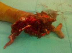
BEWARE! Gory Images

Another case of firecracker associated injuries here.

firecracker1

firecracker2

For those who don’t believe in that this kind of accidents really happen, this is a story who GruntDoc wrote, with the same outcome.

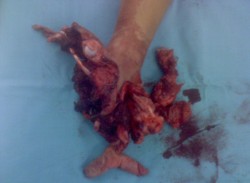
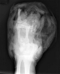
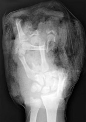
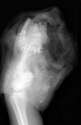
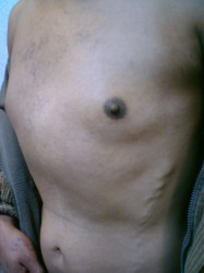
The pictures are self explaining.

GruntDoc says: “The devastation on this X-ray is nothing compared to the actual flesh-and-blood injury. A life changed in a second.”

Why this Spam?

Constantly i’m receiving comments to buy some drugs via online, is there a way to stop this? Is there a regulation? I’m getting every week a post with this words on it:

viagra, xanax, XanaX, online alprazolam, Valium, Toradol, morphine, ultram, blackjack, phentermine, poker, Online Casino, Levitra, Fioricet, illegal drugs

Is there any kind of law to stop illegal promotion of this kinds of medications? I’m sick of it.

Regards,
Jon Mikel Iñarritu, M.D.

Getting NOT enough or TOO MUCH sleep increases risk of type 2 Diabetes Mellitus

Via Diabetes Care Journal.

A cohort study (made by Yaggi, Araujo and McKinlay) who enroll men from 1987-9 to 2004 showed the next results:

Men reporting short sleep duration (< =5 and 6 h of sleep per night) were twice as likely to develop diabetes, and men reporting long sleep duration (>8 h of sleep per night) were more than three times as likely to develop diabetes over the period of follow-up.

So, If you get too much or not enough sleep, you can develop Diabetes Mellitus.

Source: Diabetes Care 2006;29:657-661.

Regards,

Jon Mikel Iñarritu, M.D.

Costochondritis (Tietze’s syndrome)

I’ve been asked –and consulted- about “rare chest pain” in several times. I could notice that people is always worried about any kind of chest pain because the fear of suffer a heart attack or a pulmonary problem. Chest pain is one of the most common symptoms that require medical attention. You –as physician- should always exclude this topics (cardiac and pulmonary) in the first place. You should keep in mind that there is a disease called costochondritis (Tietze’s syndrome) once you ruled out the main fear conditions (pulmonary and cardiac).

Costochondritis
is an inflammation of the costo-sternal joint (rib-sternum) or it could be an inflammation between the costo-chondral joint (rib-rib cartilage). The group mainly affected is that woman over 40s.

Etiology (causes)
: Direct injury to the chest, viral infections (cold / flu), idiopathic (the cause cannot be found).

Its clinical manifestations: Pain, tenderness in those joints I already mention earlier. This pain and/or tenderness get worse when you touch the involved site or move in a certain direction.
The diagnosis it’s mainly a clinical one and the physician should always exclude a heart attack and other important things.

The gold-standard of treatment is NSAIDs (non-steroid anti-inflammatory drugs like aspirin, diclofenac, naproxen, ibuprofen, acetaminophen, etc.) for one or two weeks (this disease usually lasts for this period of time). Some patients respond well to putting a local heating pad.

You have to remember that when you have chest pain, you have to look for a health care provider immediately to exclude other serious conditions.

Regards,

Jon Mikel Iñarritu, M.D.

Spanish (Español) article

Traumatic thoracic aortic rupture

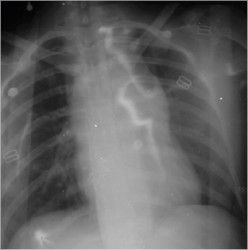
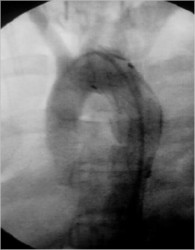
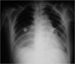
A previously healthy 17 yo woman came in to the ER because she has suffer an automovilistic accident. On the initial evaluation she was with pulse ‘parvus et tardus’ with altered mental status. A chest x-ray was ordered and found an aortic rupture.
Initial Chest X-Ray

Because the bleeding was contained and she has just a little hemodynamic instability, the choice was endovascular treatment of traumatic thoracic aortic rupture.
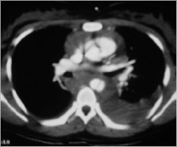
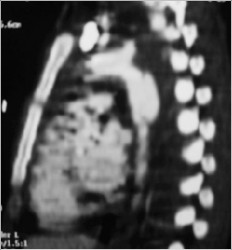
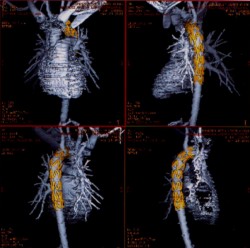
A CT Scan was ordered preoperatory:

CT Scan - Aortic Rupture

CT Scan - Aortic rupture

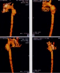
Angio TC

Endovascular Procedure

Flouroscopy - Stent

After Procedure

Reconstruction

Postoperatory Chest X-ray

The outcome was excellent and the patient is in good shape rightnow with a normal lifestyle.

Regards,

Jon Mikel Iñarritu, M.D.

Insulinoma & Whipple triad – Surgery (update)

Insulinoma

Insulinoma is the most common functioning tumor of the pancreas, and affected patients present a tableau of symptoms referable to hypoglycemia (symptoms of catecholamine release), mental confusion and obtundation. Many patients have symptoms for years. Some have been greatly troubled by emotional instability and fits of rage, often followed by somnolence.

Incidence: This tumors are so rare. Probably the best series of insulinomas is from the Olmsted County (Mayo Clinic). This study (cohort) was seen over a sixth decade period (’27 to ’86). The incidence there was 0.4 per 100,000 person-years (or four cases per million per year).

The diagnostic hallmark of the syndrome is the so-called Whipple triad, wich consists in symptoms of hypoglycemia (catecholamine release), low blood glucose level (40 to 50 mg/dL), and relief of symptoms after intravenous administration of glucose. The triad is not entirely diagnostic, because it may be emulated by factitious administration of hypoglycemic agents, by rare soft tissue tumors, or occasionally by reactive hypoglycemia. The clinical syndrome of hyperinsulinism may follow one of two patterns or sometimes a combination of both.

The symptom complex may be due to autonomic nervous overactivity, expressed by fatigue, weakness, fearfulness, hunger, tremor, sweating, and tachycardia, or alternatively, a central nervous system disturbance with apathy (or irritability or anxiety), confusion, excitement, loss of orientation, blurring of vision, delirium, stupor, coma, or convulsions.

Because cerebral tissue metabolize only glucose, prolonged profound hypoglycemia may cause permanent brain damage. Clinicians need to be alert to this when attempting to induce hypoglycemia by fasting.
Preoperative NPO orders must be accompanied by intravenous administration of glucose.

The diagnosis: Is established by demonstrating inappropriately high serum insulin concentrations during a spontaneous or induced episode of hypoglycemia. Then imaging techniques are used to localize the tumor. The available procedures include spiral CT, arteriography, ultrasonography (transabdominal and endoscopic), and 111-In-pentetreotide imaging. Transabdominal ultrasonography is our preferred initial test.

Insulinomas are small (usually smaller than 1.5 cm), usually single (only 10% are multiple and those are usually associated with MEN 1 syndrome), usually benign (only 5% to 10% are malignant), and usually hard to find. Success in localization often parallels the degree of invasiveness of the study.

Its treatment:
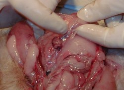
Treatment for insulinoma is surgical. At operation, the incision is dictated by operator preference, either a midline incision from the xiphoid to below the umbilicus or a bilateral subcostal incision. Exposure should be generous, and mechanical ring retractors are an asset. The entire abdomen should be explored, with particular attention being paid to possible liver metastases.

The head of the pancreas should be palpated carefully and examined anteriorly and posteriorly; the body and tail of the pancreas should be palpated, dividing any ligamentous attachments to the spleen, delivering the spleen into the wound, and rotating the tail anteriorly to allow palpation and visualization.

Anyone operating patients with pancreatic islet adenomas should be familiar with techniques for, and limitations of, intraoperative ultrasonography. The higher-resolution (7.5-to 10-MHz) transducers are used in the pancreas; because of its greater depth of penetration. Islet tumors are detected as sonolucent masses, usually of uniform consistency. Several reports attest to the high degree of accuracy of intraoperative ultrasound.

Review of current literature leads to the almost startling conclusion that nearly all syndromes of hyperinsulinism are due to insulinomas and that nearly all insulinomas can now be detected before or during surgery. Having said this, there appears to be scant justification for an empirical (“blind”) partial pancreatectomy except in patients with insulinoma plus MEN 1.

Most insulinomas are benign and can be enucleated. Nutrient vessels in the bed of the adenoma should be cauterized.

The 10% of patients with hyperinsulinism who have MEN 1 syndrome have multiple islet tumors, one of which is usually dominant and responsible for the excessive insulin output.
Insulinoma