Updating site

In the next couple of weeks I will be updating this site (style, layout, design, features).

From time to time there could be errors on displaying its new features or contents.

So please feel free to report any inconvenience when you are browsing Unbounded Medicine.

Thanks.

Update 07/24/06: While we finish the design, I will be posting on regular basis.

Laparoscopic vs Conventional Nissen fundoplication

From Annals of Surgery

A randomized trial that compares (subjectively and objectively) the laparoscopic versus conventional Nissen fundoplication in 5 years.

The comparision was made with 148 (79 laparoscopic vs 69 patients who were requested to fill in a questionnarie and to undergo esophageal manometry and 24 hours pH-metry.

Results: At 5 years follow-up, 20 patients had undergone reoperation: 12 after laparoscopy (15%) and 8 after conventional (12%). There was no difference in subjective outcome, with overall satisfaction rates of 88% (lap) and 90% (conv). Total esophageal acid exposure times (pH < 4) were 2.1% +/- 0.5% and 2.0% +/- 0.6%, respectively (P = 0.21). Antisecretory medication was taken daily in 14% and 16%, respectively (P = 0.29). There was no correlation between medication use and acid exposure and indices of symptom-reflux association. No significant differences between subjective and objective results at 3 to 6 months and results obtained at 5 years after surgery were found.

It concludes that the effects of laparoscopic and conventional are sustained up to 5 years and the long-term results are comparable. A substantial minority of patients in both groups had a second antireflux operation or took antisecretory drugs, although the use of those medications did not appear to be related to abnormal esophageal acid exposure.

I think laparoscopic approach is clearly superior due to the recuded hospital days and short convalescence period.
Regards,
Dr. Jon Mikel Iñarritu

Migraine in women is linked with cardiovascular disease

Via JAMA. 2006;296:283-291.

Migraine with aura is associated with increased risk of major cardiovascular disease (CVD), myocardial infarction, ischemic stroke, and death due to ischemic CVD, coronary revascularization and angina. Active migraine without aura was not associated with increased risk of any CVD event.

This prospective cohort-type study enrolled 27,840 women aged 45 years or older who were participating in the Women’s Health Study, were free of CVD and angina at study entry (1992-1995), and who had information on self-reported migraine and aura status, and lipid measurements. This report is based on follow-up data through March 2004.

At baseline, 5125 women (18.4%) reported any history of migraine; of the 3610 with active migraine (migraine in the prior year), 1434 (39.7%) indicated aura symptoms.

During a mean of 10 years of follow-up, 580 major CVD events occurred. Compared with women with no migraine history, women who reported active migraine with aura had multivariable-adjusted hazard ratios of 2.15 (95% confidence interval [CI], 1.58-2.92; P<.001) for major CVD, 1.91 (95% CI, 1.17-3.10; P = .01) for ischemic stroke, 2.08 (95% CI, 1.30-3.31; P = .002) for myocardial infarction, 1.74 (95% CI, 1.23-2.46; P = .002) for coronary revascularization, 1.71 (95% CI, 1.16-2.53; P = .007) for angina, and 2.33 (95% CI, 1.21-4.51; P = .01) for ischemic CVD death.

After adjusting for age, there were 18 additional major CVD events attributable to migraine with aura per 10 000 women per year. Women who reported active migraine without aura did not have increased risk of any vascular events or angina.

It will be nice when a novel study evaluates the prevention (triptans, ASA, beta-blockers) of this association between migraine with aura and CVD.

Link to: Acetaminophen + aspirin + caffeine to treat acute attacks of migraine

Regards,

Jon Mikel Iñarritu, M.D.

Intussusception

Intussusception is the invagination of a part of the intestine into itself, in other words is the prolapse of one part of the intestine into the lumen of an immediately adjoining part. It is the most common abdominal emergency in early childhood.

Epidemiology: Most episodes of intussusception occur in otherwise healthy and well-nourished children. Approximately 60% of children are younger than one year old, and 80 percent are younger than two. Is the most common cause of intestinal obstruction in children between 3 months and 6 years old. It appears to have a slight male predominance with a male:female ratio of approximately 3:2.

Etiology: The vast majority of cases is unknow. Another causes are Meckel’s diverticulum, polips, intestinal tumors, Henoch-Schönlein purpura, strange bodies, etc.
The most common form of this disease is the ileo-colic and ileo-ileo-colic invaginations. It could be associated with the rotavirus vaccine.

Clinical picture: Acute onset of intermittent, severe, crampy, progressive abdominal pain, accompanied by inconsolable crying and drawing up of the legs toward the abdomen. This painfull episodes occurs at 15 – 20 minute intervals and then become more frequent and severe. Vomiting may follow episodes of abdominal pain.

This episodes can be followed by vomiting and the passage of “currant jelly” stool (a mixture of blood and mucous). A sausage-shaped abdominal mass may be felt in the right side of abdomen. The prevalence of blood in the stool is as high as 70% if occult blood is included.

Diagnosis: It is based on index of suspicion, frequently the diagnosis is stablished with contrast studies (wich could be also therapeutic).

The abdominal plain film (see below) may be helpful because they may show frank intestinal obstruction or massively distended loops of bowel with absence of colonic gas.
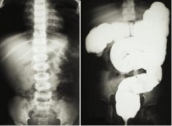
Intussusception. Plain film and barium enema

The ultrasound can be useful also, with a sensitivity and specificity approach 100%. The classic finding is a “bull’s eye” or “coiled spring” lesion (see below) representing layers of the intestine within the intestine.
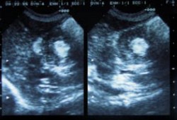
Intussusception. Ultrasound

Treatment: Nonoperative reduction using barium or air contrast techniques is successful in approximately in 75 – 90% (in the first 24 hours) of patients with ileo-colic intussusception.
Surgery is indicated when nonoperative reduction is incomplete or when a persistent filling defect, indicating a mass lesion is noted. Broad-spectrum intravenous antibiotics should be given before surgery. Manual reduction at operation is attempted in most cases, but resection with primary anastomosis needs to be performed if manual reduction is not possible or if a lead point is seen.

Regards,
Jon Mikel Iñarritu, M.D.
Early Removal of Prophylactic Drains

From Annals of Surgery

This article reminds me the risk of intraabdominal infections with a drain left there more than 4 days on the PO.

For patients with pancreatic head resection, a drain should be removed as early as the 4th PO day. This reduce the incidence of PO intraabdominal infection.

Methods: A total of 104 consecutive patients who underwent pancreatic head resection were enrolled in this study. To assess the value of prophylactic drains, we prospectively assigned the patients into 2 groups: group I underwent resection from January 2000 to January 2002 (n = 52, drain to be removed on postoperative day eight). group II underwent resection from February 2002 to December 2004 (n = 52, drain to be removed on postoperative day 4). Postoperative complications in the 2 groups were compared.
Results: The rate of pancreatic fistula was significantly lower in group II (3.6%) than in group I (23%) (P = 0.0038). The rate of intra-abdominal infections, including intra-abdominal abscess and infected intra-abdominal collections, was significantly reduced in group II (7.7%) compared with group I (38%) (P = 0.0003). Eighteen of 52 (34.6%) patients in group I had an inserted drain beyond 8 days, whereas only 2 of 52 (3.7%) patients in group II had an inserted drain beyond 4 days (P = 0.0002). Cultures of drainage fluid were positive in 16 of 52 (30.8%) patients in group I, and in 2 of 52 (3.7%) patients in group II (P = 0.0002). Intraoperative bleeding (>1500 mL), operative time (>420 minutes, and the period of drain insertion were significant risk factors for intra-abdominal infections (P = 0.043, 0.025, 0.0003, respectively). The period of drain insertion was the only independent risk factor for intra-abdominal infections by multivariate analysis (odds ratio, 6.7).
Conclusion: Drain removal on postoperative day 4 was shown to be an independent factor in reducing the incidence of complications with pancreatic head resection, including intra-abdominal infections

Regards,
Jon Mikel Iñarritu, M.D.

Cervical Cancer Vaccine (gardasil)

FDA approves the fist vaccine for cervical cancer, a week ago, the vaccine was aprooved in Mexico. With the name of Gardasil, and a cost of US$120.00/dose (2 doses to complete the treatment), I celebrate this effort against cancer.

Gardasil

Here in Mexico, cervical cancer is by far, the most common type of cancer (24.4%) in general population (including males). In females, again is the most common type of cancer (36%). The most common risk factor is infection from human papillomavirus (HPV).

This virus causes cervical cancer, precancerous genital lesions and genital warts; the vaccine prevents against HPV types 16, 18 (causes 70% of cervical cancer cases); 6 and 11 (causes genital warts).

Let’s celebrate this fight against cancer.

Regards,
Jon Mikel Iñarritu, M.D.