Being Curious – Doctor’s Bag

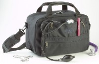
doctor's bag

I was reading at Patient Plus an article of what to put in the Doctor’s Bag and I wonder:

In the past, the vast majority of doctors used a Doctor’s Bag, in fact there are museums with different kinds of Doctor’s Bags… but things are changing.

Some physicians still use a Doctor’s Bag to deal with all the medical stuff like stethoscope, diagnostic sets, aneroid sphygmomanometer, thermometer, drugs and depending on your speciality or practice, other usefull things.

Do you carry a doctor’s bag?

What do you have in there?

If you don’t carry a bag, what do you carry allways (mobile phone, PDA, lab coat, scrubs, handbooks)?

Thank you for your answers and regards,

Dr. Jon Mikel Iñarritu

Gallstone Ileus

Gallstone Ileus is an infrequent cause of mechanical bowel obstruction. It is caused by an impaction of a gallstone in the terminal ileum by passing through a billiary-enteric fistula (often from duodenum). It occurs more frequently in women with average age of 70 years.

Clinical picture: Episodic subacute obstruction in an elderly female. Abdominal pain and vomiting which subside as the gallstone becomes disimpacted, and only recurs again as the progressively larger stone lodges in the more distal bowel lumen. Intermittent symptoms may be present for some days prior to evaluation. Hematemesis could occur as an occasional complication that is due to hemorrhage at the site of the biliary enteric fistula.

Physical examination: The patient may be febrile and often appears dehydrated. Common abdominal signs include distension and increased bowel sounds. Jaundice is uncommon, occurring in less than 15% of cases. Many affected patients have serious concomitant medical illnesses, including coronary disease, diabetes mellitus or pulmonary disease.

Diagnosis: The most important diagnostic test is abdominal plain film. The diagnosis of gallstone ileus is made preoperatively in about one-half of cases. The radiographig findings of gallstone ileus, are:

  • Signs of partial or complete intestinal obstruction
  • Air in the biliary tree (pneumobilia)
  • Direct visualization of the stone
  • Change in position of a previously located stone
  • Two adjacent small bowel air-fluid levels in the right upper quadrant

Treatment: First of all, as any kind of bowel obstruction, the objective is to relief intestinal obstruction after adequate fluid repletion. The options are enterolithotomy, cholecystectomy, and fistula division, with or without common bile duct exploration (one-stage procedure), with definitive repair performed at a second operation (two-stage procedure).
The treatment of choice is the enterolithotomy wich consits in localize and extract the gallstone. Often the cholecystectomy is contraindicated by comorbidities and the general state of the patient.

Case presentation: A 72 year old woman with heart faliure, hypertension and diabetes came to the office with a chief complaint of chills, abdominal pain, nausea and vomiting followed by inability to pass flatus 8 hours prior to consult.

On physical examination the patient was febrile and appeared dehydrated. Abdominal exam: distension and increased bowel sounds. No jaundice.

Labs: Hyperglicemia, mild renal failure, hypernatremia and leukocytosis.

Radiographic tests: Abdominal plain film with signs of bowel obstruction and with not apparent cause, negative US. CT scan just with signs of intestinal obstruction, not pneumobilia, not obstructing gallstones.

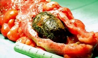
The patient bacame hemodinamically unstable while performing the US and we decided to open her. On the OR we found the level of obstruction at terminal ileum and we felt a solid mass. We performed an enterolithotomy and the result was this:

Gallstone Ileus

Regards,

Dr. Jon Mikel Iñarritu

Splenic Injury and Hemoperitoneum in Blunt Trauma

The following pictures are from different clinical cases.

In Blunt Trauma the spleen and the liver are injuried in 40 and 20 percent, respectively.

In the ER, you have three four options in the Work-Up of a patient with blunt abdominal trauma

  • Peritoneal Lavage: Now just performed if you haven’t access to ultrasound (lack of money or lack of trained personnel). It’s indicated in specific cases, as in patients with cranial trauma, spinal trauma, for example.

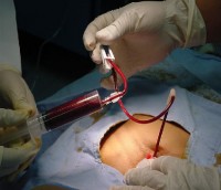
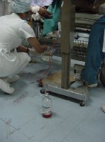
Positive Peritoneal Lavage - 1 Positive Peritoneal Lavage - 2

Look the blood in the syringe and the tube. This is a positive peritoneal lavage due to blunt trauma.
  • Ultrasound (a.k.a. FAST or Focused Abdominal Ultrasound for Trauma): This is one of the most used techniques, and should be performed for those patients hemodynamically unstables who can’t go to the coputed tomography room.

Positive FAST
Look the irregular border. This is a positive FAST for hemoperitoneum due to blunt trauma.
  • Computed Tomography: This technique has a magnific resolution for spleen, liver and vessels. Excellent evaluation for retroperitoneum. It has one inconvenience, hemodinamically unstable patients are not candidates for this analysis.

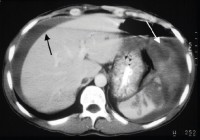
Spleen Injury and Hemoperitoneum
Black arrow = Hemoperitoneum.
White arrow = Injuried spleen
  • Diagnostic Laparoscopy: Almost never recquired.

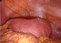
Positive Diagnostic Laparoscopy
Look the blood in the abdominal cavity, this is a positive Laparoscopy for hemoperitoneum due to blunt trauma.

The controversy is when the hospital is not capable of performing the standarized protocol in blunt abdominal trauma (economic reasons), we doctors have to adequate to the circumstances and do our best effort.

For example, the first two photographs (peritoneal lavage) were taken at the Mexican Red Cross where money is the big problem. The rest, were taken at the ABC Medical Center, a nice private hospital. This are the contrasts of healthcare system and medicine practice in developing countries.

Regards,

Dr. Jon Mikel Iñarritu

New ePOCRATeS

With new look, new content and new features appears the new ePOCRATeS Rx.

I’ve been trying this brand new version of ePOCRATeS and I think that it’s quite better than the previous version.

The main features are:

  • My Epocrates personalized homepage
    • Go straight to the drug monographs you recently viewed
    • Create shortcuts to get to your favorite applications faster
    • Launch MobileCME activities in your specialty
    • Edit your profile
My ePOCRATes My ePOCRATeS News and updates Lookup
  • New drug safety content
    • Therapeutic Drug Levels
    • Monitoring Parameters
    • Non-Interchangeable Forms
    • Pregnancy and lactation information
  • New decision support tools
    • Corticosteroid Converter
    • Narcotic Analgesic Converter
    • Heparin Dosing Protocol
    • INR Calc
    • Topical Corticosteroids
  • Memory Card support (this is nice)
  • More than 3,300 brand and generic drug monographs, searchable by name or class
  • Peer-reviewed drug content summarized from a wide range of authoritative sources
  • MultiCheck drug-interaction checker (30 drugs simultaneously)
  • Integrated health insurance formularies
  • MedMath* medical calculators, e.g., Body Mass Index, Creatinine Clearance
  • Ability to personalize the application to suit your needs
  • DocAlert messaging — relevant, timely clinical and specialty news
  • AutoUpdate

Give it a try.

Regards,

Jon Mikel Iñarritu, M.D.

Mobile Data Visualization

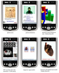
At Quinn Lab, San Diego Supercomputer Center, doctor Quinn, has an outstanding project called Patient Notes with mobile devices.

The famous and enthusiastic project of Dr. Quinn and Dr. Wright, consists in the transmission of medical data to mobile devices such as PDAs and cell phones to enable medical workers in the field to instantaneously gain access to, view and prognosticate on complex medical visualizations.

With this software, medical notes and patient tests can be downloaded onto a cell phone or PDA in just minutes.

All the data on a phone is stored in the memory expansion slot. In these medical phones, however, Instead of music and digital pictures, it could hold a virtual scan of the body and much more.

Examples:

Dr. Quinn's Patient Notes
The 3-D mobile medical data program should be available within a year.

This is a good start to revolutionize the way of getting access to medical information about our patients.
Its uses are endless: ambulance, emergency departments, office, etc.

Google Health Co-op

Co-op

If you consider the info annotated here is useful, please add me to your trusted annotators at Google Co-op.

This program of Google, improves your health and medicine searches and its objective is to help people to find medicine and health related topics. The fact is that most of the people gets here, use google as a search engine.

If you are participating in Google Co-op (specially in Health), please let me know so we can exchange subscriptions to our trusted content. So if you like the quality of the information I post, please subscribe to my Co-op here

I would like to thank you for your support.

Regards,
Jon Mikel Iñarritu, M.D.

Worst case of hemorrhoidal prolapse ever & PPH

BEWARE! Gory image

A 45 year old male with history of chronic reducible hemorrhoidal disease came to the emergency department with excruciating pain, rectal bleeding and an anal non-redicible mass after a bowel movement.

On examination, we found this:
hemorrhoidal prolapse

In this case, the only way to reduce the prolapse is a procedure for prolapsing hemorrhoids (PPH) which is an innovative approach to the management of enlarged, prolapsing hemorrhoids. Rather than rely on excision of the complexes, the primary goal is to reduce the hemorrhoidal tissue and anoderm to the correct anatomical location within the anal canal. It is important to keep this goal in mind, because the misinterpretation of anodermal migration externally as external hemorrhoids leads to unnecessary excision of sensate skin during PPH. In fact, with a correctly performed procedure, the relocated anoderm will shrink over time.

For this, we need an hemorroidal circular stapler kit which includes: clear plastic anoscope, half-slit anoscope, suture threader, and the 33 mm stapler.

Here is an animation video of how the procedure works, and how does the anal canal looks like before and after the procedure.

Regards,

Jon Mikel Iñarritu, M.D.

More information of hemorrhoidal disease in this site is here:
Thrombosed Hemorrhoid

McGraw-Hill’s Access Surgery

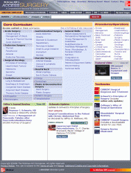
With a “very cheap” subscription rate of $995 (OMG!!) you can have access to Access Surgery, inside you can find this stuff:

  • Surgical Videos: Columbia University College of Physicians and Surgeons Surgical
  • Animations: Adapted from Zollinger’s Atlas of Surgical Operations, 8th Edition
  • Textbook: Schwartz’s Principles of Surgery, 8th Edition
  • Textbook: CURRENT Surgical Diagnosis & Treatment, 12th Edition
  • Surgical Atlas: Zollinger’s Atlas of Surgical Operations, 8th Edition
  • Surgical Anatomy: Skandalakis’ Surgical Anatomy
  • Board Review: Surgery Review Illustrated
  • Quick Answers: CURRENT Consult Surgery Table of contents (see the image below).

AccessSurgery

Special Features:

  • Create your own Board Review test from Surgery Review Illustrated, then email results to your program director
  • View Surgical Videos on your iPod from the Schwartz editorial team
  • Look up a symptom or disease in our Differential Diagnosis (DDx) tool
  • Download the free PDA diagnostic tool from CURRENT Consult Surgery
  • Assess the extent of disease through Cancer Staging Tables
  • Discover related Government Guidelines by clicking a pre-populated search box
  • Launch a Customized Search for just videos, just images, or view full-site results
  • Research medication indications, dosages, and contraindications through an integrated Drug Database provided by Gold Standard.

The site was launched on July 18th.

Will you pay for this material? (It’s a question, because I don’t)

Regards,

Jon Mikel Iñarritu, M.D.